

Scientists in Germany believe they have found a solution for people suffering from retinitis pigmentosa (RP) and macular degeneration (MD), as was explained in last week’s best-read story. RP is a congenital eye disorder that occurs in people between the ages of 30 and 50. It starts with night blindness and a narrowing field of vision, the vision gradually continues to deteriorate and this may even lead to blindness. With macular degeneration, the macula (yellow spot) in the retina deteriorates. This causes sufferers to see less sharply in the center of their visual field. For example, they have difficulty reading. Usually people with MD do not go completely blind.
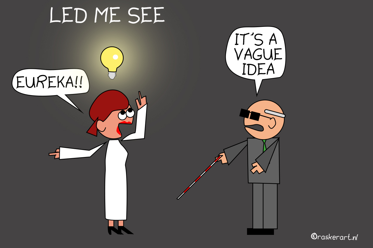
By using LED lamps that individually adjust the contrast and amount of blue in the light per patient, researchers at the Fraunhofer Application Centre Soest and the Department of Electrical Engineering at the University of Applied Sciences Südwestfalen hope to be able to improve patients’ vision.
Kamermans is a professor of neurophysiology attached to the University of Amsterdam and head of the Kamermans group at the Brain Institute where he has been doing fundamental research on the workings of the eye for 30 years. What are his views on these special LED lights?
Both disorders function differently
Firstly, he would like to point out that both eye conditions affect the retina in different ways, which means that the symptoms are also not comparable. To explain this better, here is a crash course in how the eye works: “Our eye captures the reflection of light from the object we are looking at. These particles of light enter light-sensitive cells inside the retina. This is where the cones are which are most common in the macula, the central part of the retina. These allow us to see sharply and in color during the day. To see at night or in places with relatively little light, we have rods. These are much more light-sensitive than cones and are found more towards the edge of the retina. We cannot see color with these,” explains Kamermans.

The rods in the retina slowly die off in someone with the congenital defect RP. “As a result, patients can no longer see anything in the dark and vision gets worse at the edges. It causes a kind of tunnel vision,” says the professor. Macular degeneration, he says, works exactly the other way around. In this case, the cones in the macula die off, leading to diminished sharpness of vision in the center of the visual field in patients. “As a result, they often recognize faces less quickly and have difficulty reading. This is almost impossible to treat. Patients are taught to then look at things more with the edges of their eyes. But this is not a proper solution; vision still remains blurry.”
Remains to be seen if light glasses can improve sight
He says there is a lot of experimentation going on at the moment with light glasses or special lamps like those in Germany. “Some patients like these options, but that’s very subjective, of course. Not an unimportant consideration, by any means. But there are not very many blue cones in the macula. These are more towards the edge and are more sensitive to blue light. I have yet to see if it can be objectively determined whether any improvements in vision can be made this way.”
A lot of researchers compare the workings of the retina to that of a camera. Undeservedly so, in Kamermans’ opinion. “The retina is much more complicated than people and also scientists often think. A camera relays every pixel and uses it to build up an image. Whereas the retina filters out all sorts of things before sending signals to the brain. It makes constant calculations, so to speak, and only sends on signals to the brain that deviate. It is a stream of information about movement of objects, light intensity, colors and positive or negative images.”
All of these signals reach the brain in different areas. Science has not yet figured out exactly which signals belong to which areas of the brain. This is such a complex process that Kamermans does not see retinal prostheses becoming widely used anytime soon. “These implants translate light into an electronic current that stimulates the cells of optic nerves. These currents are just not capable of stimulating specific cells, cells that say something about the shade of red or how fast something is moving, for example. This means that vision is limited and the results are disappointing. The technique has not yet been developed far enough to ensure that the right signals are sent to the right place.”
Gene therapy will lead to a breakthrough
He has more confidence in gene therapy as a way to prevent the symptoms of RP. “The innate gene causes light-sensitive pigment in a cell to fold up incorrectly and not be broken down. This causes the cell to become crammed with faulty photopigment and then eventually die off. In gene therapy, this incorrect gene is cut in half. This prevents the pigment from accumulating and the cell from dying. The research results are promising and this breakthrough is definitely going to happen.”
Another promising technique for treating RP is optogenetics. This involves influencing the behavior of a cell by inserting a light-sensitive gene. In the research by Kamermans’ group, this gene came from light-sensitive ion channels. These are developed in blue-green algae in order to detect light. “By inserting these into the retina, they replace the function of the photoreceptors that have broken down,” he goes on to explain.
The key here is to insert the gene in the right dosage and at an early stage of the disease. “The retina has a layered structure, whereby the first layer takes care of all the calculations before signals go to the brain. The disease is progressive so you have to catch it in time. Otherwise that layer is already too far damaged. It is possible to make a deeper layer sensitive to light. But that produces poorer results because you then lack those calculations from the first layer.”
Further research needed
The first three patients were treated with a relatively low dose by the French research group Gensight, Kamermans reports. ” They responded well to this. The plan is to further shape this study. But I haven’t heard anything more about this for a while because of corona – at physical conferences you chat to each other in between lectures, but this happens less online.”
Back in the Netherlands, Kamermans and his colleagues had to stop researching optogenetics because they were no longer getting enough retinas. “Previously, we received the residual material for scientific research from the cornea bank. But since this was transferred to the tissue bank, only eyes are allowed to be used for corneal research; the rest is thrown away. While we know there is an ample supply. This is extremely frustrating. We may not be working towards clinical solutions with our research at the moment, but our fundamental knowledge can definitely contribute to this.”
Related Posts


