
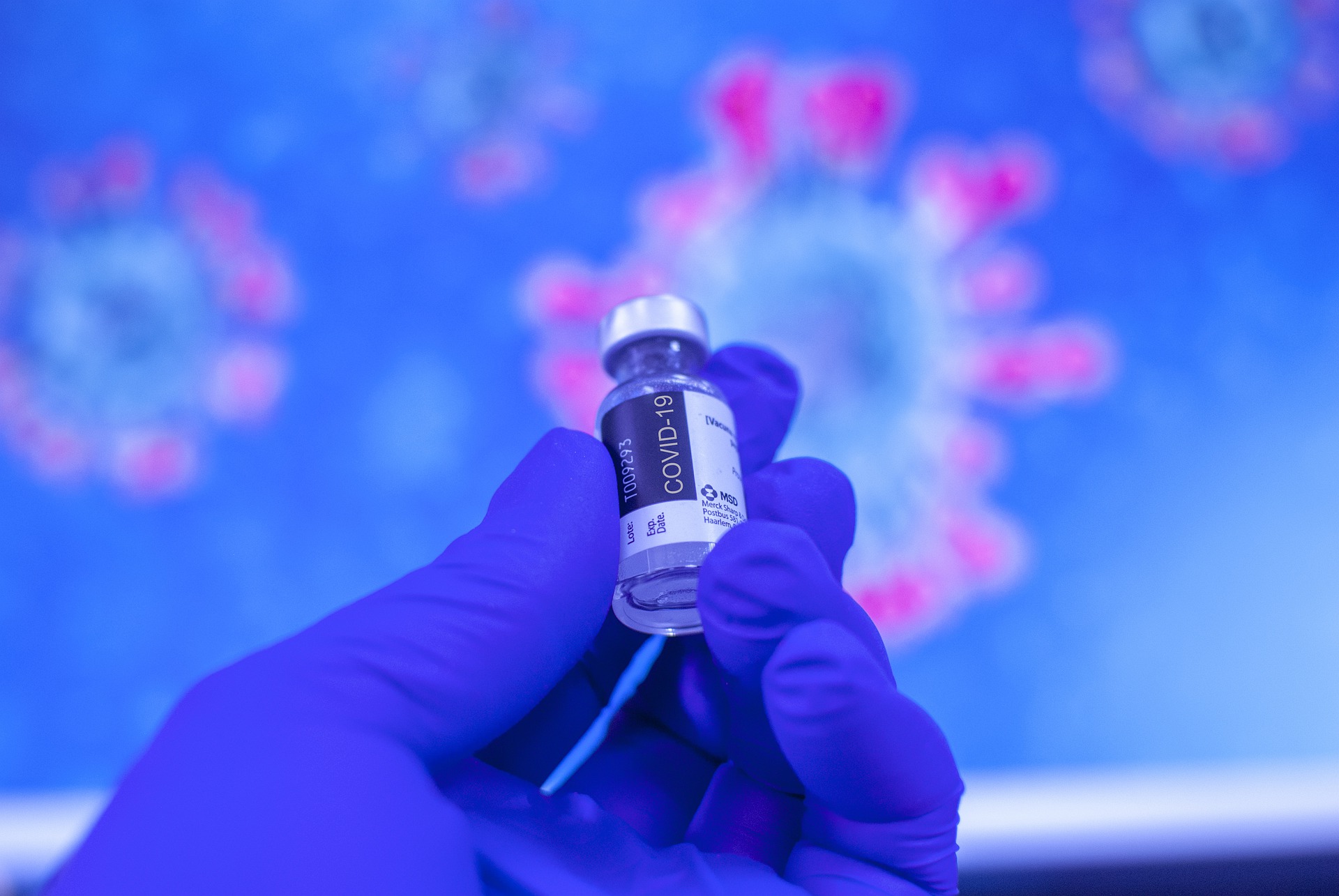

Researchers at Chalmers University of Technology, Sweden, have succeeded in developing a method to label mRNA molecules. This allows them to follow their route through cells in real time using a microscope. And this can be done without affecting their properties or their subsequent activity. This breakthrough could be of great importance for the development of new RNA-based drugs. The corona vaccines from BioNTech/Pfizer (Comirnaty) and Moderna are such examples.
RNA-based drugs offer a range of new opportunities for preventing, treating and potentially curing diseases. Currently, administration of RNA drugs into cells is inefficient. In order for new vaccines to reach their potential, administration methods need to be optimized. The Swedish researchers have published a new method in the prestigious Journal of the American Chemical Society. This could push the development of drugs based on mRNA a huge step forward.
“Our method can help solve one of the biggest problems in the discovery and development of drugs. This will facilitate the shift from traditional drugs to RNA-based therapeutics,” said Marcus Wilhelmsson, professor in the Department of Chemistry and Chemical Engineering at Chalmers University of Technology.
Fluorescent mRNA
The research behind the method was conducted in conjunction with chemists and biologists from Chalmers and the biopharmaceutical company AstraZeneca in their joint research center FoRmulaEx. A research group from the Pasteur Institute in Paris also took part.
One of the building blocks of RNA has been replaced by a fluorescent variant. It retains its natural properties. This marks a breakthrough that has never before been successfully achieved. The fluorescence also enables researchers to track functional mRNA molecules in real time and with the aid of a microscope see how they are absorbed into cells.
One challenge in working with mRNA is that the molecules are very large and charged but at the same time also quite fragile. They are unable to enter cells directly. Which is why they need to be packaged. So far, the method that has proven most successful uses very small droplets –lipid nanoparticles– to encapsulate the mRNA.
Development of new and more efficient lipid nanoparticles is still greatly needed. This is something that Chalmers researchers are also working on. To do this, it is imperative to understand how mRNA is assimilated into cells. Being able to track in real time how lipid nanoparticles and mRNA spread through cells is therefore an important asset. “The major advantage of this method is that we can easily see now where the administered mRNA goes in the cell and in which cells protein is formed, without losing the natural ability of RNA to translate into proteins,” scientist Elin Esbjörner explains.
Crucial information
Researchers in this field are able to use the method to gain more knowledge about how the absorption process works. This can speed up the discovery process of new drugs. The new method provides more accurate and detailed information than current methods of studying RNA under a microscope.
“Up until now, it has not been possible to measure the natural speed and efficiency of the way RNA works inside cells. This means that you get the wrong answers to the questions you ask when you’re trying to develop a new drug,” Marcus Wilhelmsson says. “That makes it tricky to develop a new drug.”
Read the article here in the Journal of the American Chemical Society.
Also interesting: MRNA technology can revolutionize the fight against cancer and other diseases
Related Posts


