
His limitless interest in science, data and technology led him to come up with innovations. And he may well now be holding the ‘Holy Grail of vascular surgery in his hands. Richard Lopata wants to see what’s happening to your organs and arteries under your skin. What he wants to do is visualize that for better diagnoses of, say, a narrowing in the carotid arteries or the leg. Richard Lopata is associate professor at the Eindhoven University of Technology (TU/e) research group Photoacoustics & Ultrasound Laboratory Eindhoven (Department of Biomedical Engineering) where he heads the PULS/e lab.
Technical and medical
He made it to the finals of the Chemistry Olympiad as a teenager. “I was actually always destined to do something with chemical engineering,” he adds. Chemistry was not the only thing he excelled at. As a child, he tinkered and played a lot with Lego, he always had to make something. During high school, he also “started doing a little bit of programming.” Lopato was unable to choose between the technical disciplines. “I liked all four of chemistry, physics, electrical engineering and mathematics just as much.” Which is why he went on to attend the four technical universities in the Netherlands, although he did miss the medical side. “Not so much doing surgery myself, but helping doctors make better diagnoses.”
The ideal combination turned out to be BioMedical Engineering at TU/e. A program that had just started up. He was part of the second intake and it was there that he came into contact with imaging techniques (X-ray (CT), magnetic fields (MRI) and ultrasound), signal analysis and image analysis. Subjects he is still involved with today. In the first part of his studies, it seemed that chemistry would continue to dominate, but by his own admission, he slowly strayed from that – “to things that were really new to me.”
Contrast agent
Lopata combines things. Not just technologies, but modeling physiological processes and the depiction of them. “I have never focused on one thing. Take my thesis project, for example. Research on how to use an MRI scan to make images of tumors. I was not concerned with getting the best possible picture, but rather with determining if a good amount of blood was flowing through a tumor.”

He used a contrast agent that was injected into the patient. That substance then slowly leaked into the tumor. “With mathematical models of the exchange of those particles, you were able to model the leakage process. That way, you could see how terminal or leaky the vessels of such a tumor were. You can often see in malignant cells that there is a dead core.”
For his PhD research, Lopata found a placement at Radboudumc in Nijmegen, where ‘his ultrasound career’ kicked off. “I switched from MRI to ultrasound there,” he says. While there, he did research on how well the heart muscle can contract and made 2D and 3D images of the deformations of the heart. He then went on to Maastricht where he conducted research on aneurysms, as in dilations of part of the vascular system.
Risk of rupture
“You can have an aneurysm for years without noticing it. You don’t notice it until it’s too late – that’s when such an aneurysm ruptures. Then you have a massive hemorrhage; this is fatal in 75 to 95 percent of cases.”
“In many cases, an aneurysm like this is detected because, for example, someone has fallen or has problems with their heart. Then a screening process follows. If it is 5.5 centimeters in diameter in men and 5 centimeters in women, then you get a stent.”
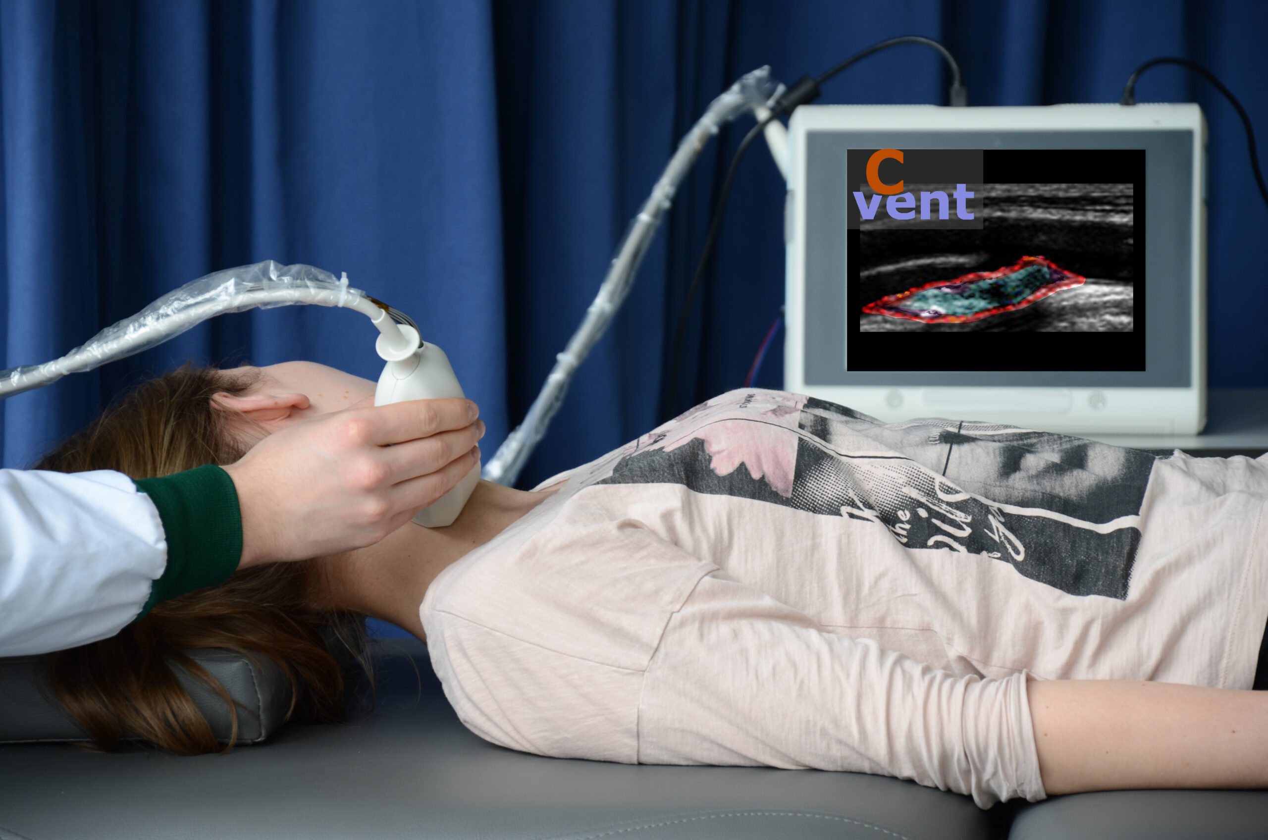
At present, that is the only measure to assess the risk of rupture, Lopata explains. “There’s simply not much more that you can say about that kind of dilation .” More can be seen with a CT scan though. But to be able to give all patients such a scan several times a year is far too expensive, Lopata explains. Also, a CT scan releases radiation and uses a contrast agent that in turn is bad for the kidneys, the researcher adds. ” This is why we explored in Maastricht, and later in Eindhoven (Catharina Hospital), what ultrasound could do to enable doctors to make better clinical decisions.”
Imaging technology specialists and physicians responded that ultrasound would not be good enough to image an aneurysm. Lopata acknowledges that with an ultrasound scan, you can only scan a small part, while with a CT scan you can make images of the whole person. Also, the contrast level on the images is much less than that of CT, the researcher notes.
Patient-specific
“With ultrasound, you can’t distinguish between two different tissues as clearly. On the other hand, the resolution of ultrasound (ultrasound) is a lot higher and we can make images very quickly in terms of time. Also, we can look at a heart or aneurysm, for example, from all directions.”
Lopata saw opportunities for using ultrasound to scan images of the stiffness and elasticity of such an aneurysm in the aorta. “By doing that, you get much more patient-specific information. Blood pressure and elasticity provide a certain degree of tension and stretch in the wall. Those can both be a measure of the likelihood of rupture.” Lopata combined knowledge about deformation and strain measurements of the heart to the stretchability of the aorta. “It was a very challenging study, but it succeeded.”
Biomechanics (Department of Biomedical Engineering), where he continued his research on fragile arteries with the Catharina Hospital, among others. He set up the Photoacoustics and Ultrasound Laboratory Eindhoven, PULS/e in 2014. In 2017, he was appointed senior lecturer at the university. PULS/e became an independent research group in 2021. For his research, Lopata secured several European and national grants, which he has used to continue his research on aneurysms and stenosis in blood vessels, among other things.
Holy Grail
An operation on, for example, a narrowing in the carotid artery is extremely invasive. Lopata: “In the Netherlands, patients with a narrowing – atherosclerotic plaque – of 70 percent or more are routinely operated on. Because of the risk of a cerebral infarction. Then a surgeon removes the inner layer of the vessel wall. “They then shut down the blood supply of your brain on one side. It is a dangerous surgical procedure with the risk of complications and sometimes a long rehabilitation period.” Not all surgeries are needed. For example, it turns out in retrospect that only 1 in 6 plaques was potentially a vulnerable, and therefore dangerous, plaque, Lopata says. “And statistically, you need to operate on one in 21 patients to prevent 1 cerebral infarction.”
Lopata works with new imaging techniques. Together with fellow researcher Min Wu, he combines sound – ultrasound – and light – photoacoustics. “Photoacoustics is now often used for basic research. The combination of sound and light opens up ways to image different tissues in the body separately from each other in a safe way. You can make the most fantastic pictures with it. What we and a number of other groups around the world are trying to do is really make this suitable for clinical imaging.”

The first integrated photoacoustic ‘probe’, an ultrasound head that measures by means of light and sound, was developed in collaboration with Catharina Hospital. Lopata, together with co-researcher and vascular surgeon Marc van Sambeek, ran a small pilot study at Catharina Hospital on patients who were to undergo surgery on their carotid artery. The study is still ongoing. But if it proves to work, then the researchers may very well have the ‘Holy Grail’ of vascular surgery in their hands.
Reflections
The research laboratory of Lopata is located on the campus of TU/e. One big difference with a university hospital is that no patients and doctors are walking around there. That difference is compensated through the collaboration with the Medical Center and Catharina Hospital. ” With the Catharina Hospital, we started one of the largest aneurysm studies within the Netherlands. That was already huge, with 350 patients, and is only set to expand further. In the meantime, there are more than 540 patients. Two doctoral students obtained their PhDs on that subject in the beginning. Now there are as many as eight PhD students running around busy with both the ultrasound side and the modeling side. What’s more, a company like Philips has also expressed interest.”
“One of the innovations of the past year is multi-perspective ultrasound. Normally you have one ultrasound probe which you hold in your own hand yourself. That’s used to transmit the sound into the body. That comes back and from those reflections we make images. We position two of those probes, and hopefully in the future more, or a whole mat, on the abdomen. One sends the sound, the other receives the reflections of that sound. If you approach the heart or an aorta from two directions, then the contrast and resolution become many times higher.”
With a conventional ultrasound machine, Lopata is now able to image an aorta at a rate of 10 3D images per second. “Compared to CT, of course, that is still not very fast, but it is radiation-free. We have now expanded it to 100 images per second. This lets us measure elasticity more accurately. By using multiple probes, we also increase the area that we can image.” Clinicians at Catharina Hospital are now using the multiple-probe method in heart and aneurysm patients as part of a new study that is being supported by TU/e and Philips.
Wearables
“On the one hand, we are making the ultrasounds more complicated with advanced techniques. On the other hand, these techniques serve as a basis for turning them into wearables. Wouldn’t it be great if a patient could wear a vest with ultrasound probes in it? Which track everything, while walking, shopping and exercising.”
Lopata is also back to working on the heart with this new technique. His ambition is to make the technique ‘wearable.’ “Because in heart patients, you are missing a lot of data as it isy. You see someone in the clinic, but those visits are snapshots. One heart patient can just walk up the stairs while another one cannot. Why is that? Or maybe later also with pregnant women, if there are complications. Then you could continuously monitor the contraction of the uterus, the heart rate and the development of the baby.”
Lopata is also working with the spin-off company Usono. This company is specialised in attaching ultrasound scanners to the body for continuous and dynamic monitoring.
Read previous articles about Usono at IO here
To the sports field
Ultrasound is harmless and inexpensive, Lopata points out. “It is my vision for the future that more and more, we will be able to replace CT and MRI scanners with sound and light. And that we can take this advanced technology from the hospital to the GP, the home or even to the sports field. That’s what we’re trying to get funding for.”
Back in 2014, Lopata mapped out several lines of research for the following 15 years. He is now halfway through. “I am immensely proud that we are able to realize our vision. That we deliver on the things that we promise. I don’t stray too much from that. Obviously, I do make use of new developments, such as deep learning. That is now seeping into my group as well. But the laws of physics also continue to apply. We look at things from that perspective: where can Artificial Intelligence (AI) help? We don’t have to use it unless it actually adds something.”
This article is part of the “This is how innovation works” series on innovation leaders. You can find the other stories here.
Collaboration
Related Posts


