
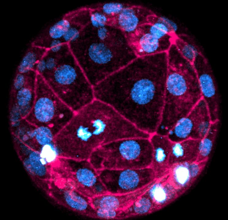
Utilising fluorescent dyes and laser microscopes, researchers from the University of Pennsylvania in Philadelphia observed real-time cell divisions and chromosome segregations, with no need for genetic alterations – thus sidestepping ethical issues. The study also uncovered crucial differences between human and mouse embryo development, underlining the uniqueness of human embryogenesis. The observations could be instrumental in developing non-invasive screening methods for embryos in IVF treatments.
The technique, which captured even chromosome segregation defects, has potential clinical applications in monitoring and selecting embryos before IVF implantation. Researchers plan to refine the technique further by using lower-intensity laser microscopes and additional dyes for longer periods.
- Scientists have achieved a groundbreaking feat by imaging human embryos at the highest-ever resolution, observing real-time cell divisions and chromosome segregations without genetic alterations;
- The study reveals crucial differences between human and mouse embryo development, highlighting the uniqueness of human embryogenesis.
- The technique has potential clinical applications in monitoring and selecting embryos for IVF implantation. It could lead to the development of non-invasive screening methods for embryos, improving success rates in IVF treatments.
Unveiling the Hidden Intricacies of Embryonic Development
The groundbreaking study, published in the journal Cell, has managed to record the first fourty hours of human embryonic development with an unrivalled level of detail. By applying fluorescent dyes, SPY650-DNA and SPY555-actin, the researchers successfully labelled genomic DNA and a protein called F-actin respectively. These labels made it possible to observe cellular events such as cell division and chromosome segregations in real time. This level of monitoring has never been achieved before and it offers a deeper understanding of early human development.
In a departure from previous approaches, this non-invasive imaging technique does not require any genetic alteration of the embryos. This addresses a major ethical concern in the field of embryonic studies, as it bypasses the need for genetic manipulation that could potentially harm the embryo. It also opens up new possibilities for studying embryonic development without ethical constraints.

Discovering the Minute Details of Human Embryogenesis
One of the significant findings of the study was the observation that cells in the outer layer of the embryo, known as the trophectoderm, lose some DNA during the interphase stage of cell replication. These errors in cell replication could be linked to chromosomal abnormalities such as aneuploidy, which is associated with pregnancy loss and implantation failure. With this new knowledge, there is potential for interventions that could correct these issues, leading to improved success rates in IVF treatments.
The researchers also discovered key differences in the early development stages between human and mouse embryos. They found that the process of compaction, where cells undergo shape alterations, starts at the 12-cell stage in human embryos compared to the 8-cell stage in mice. Moreover, compaction in human embryos is more asynchronous, leading to variations in the formation of inner and outer cells. These differences underscore the uniqueness of human embryogenesis and highlight the limitations of relying solely on animal models for understanding human development.
Setting the Stage for Groundbreaking Clinical Applications
The researchers are now planning to extend their imaging process to cover longer periods of human embryonic development. They aim to use lower-intensity laser microscopes and additional dyes to label other structures such as cell membranes. This will further enhance the resolution and detail of the images, allowing the scientists to delve even deeper into the complexities of embryogenesis.
This advanced imaging technique could have significant clinical applications in the future. It could potentially allow for non-invasive monitoring of embryos in the clinic, enabling healthcare professionals to make more informed decisions about which embryos to implant during IVF procedures. By providing a clearer picture of the embryo’s development and potential chromosomal abnormalities, it could also contribute to the development of more effective screening methods for embryos conceived through IVF.
Related Posts


