

Getting the results of a blood test still requires some patience. First, it means getting to the doctor’s before breakfast and, if necessary, waiting a while. After the blood sample has been taken, it is sent to the lab and you have to wait for the result until at least the next day. But all this could be done much faster without time-consuming lab tests because the mechanical properties of cells alone can reveal what diseases a person is suffering from.
Researchers at the Max Planck Center for Physics and Medicine (MPZPM) and physicians at the University Hospital at the Friedrich Alexander University, both in Erlangen, Germany, have now taken advantage of this in their RAPID Diagnostics project. If they succeed, soon every doctor’s office will be able to analyze blood samples quickly and reliably using AI-supported processes. This will eliminate the need to send samples to laboratories.
Faster and more cost-effective
Every patient is familiar with this situation. You have a health problem, go to the doctor, describe your complaints – and then the analysis begins. In most cases, however, taking your temperature and an ECG are not enough to make a diagnosis. A detailed blood test provides much more information, so a sample is taken that is subsequently analyzed in the lab. However, this is both time-consuming and expensive. The new RAPID method is not only faster but also less expensive and just as reliable.
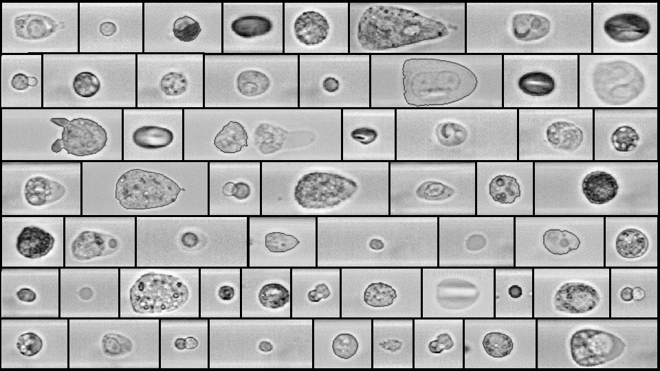
But RAPID does not stand for “fast” here, rather it is the abbreviation for “real-time analysis of physical phenotype in deformational flow.” The basis of this method is a technique in which a blood sample is sent through a transparent channel smaller than the diameter of a hair. In the process, a high-speed camera captures how the cells are deformed at about 2,000 to 4,000 photos per second. Artificial intelligence (AI) then searches these images for features that are signs of certain diseases and provides a specific suggested diagnosis. Another advantage of this method over the usual microscopic examinations is that the cells do not have to be elaborately stained in the process.
Using the method in clinical practice
The scientists and physicians led by Dr. Markéta Kubánková now want to put the RAPID technique to practical use in clinical operations over the next two years. In addition to conventional diagnostic procedures, they want to examine patient samples using the RAPID method together with the head of the laboratory at the Children’s and Adolescent Clinic, Professor Manfred Rauh, and the deputy director of the Children’s and Adolescent Clinic, Professor Markus Metzler, to create an extensive database. At the end of the project period, they plan to found a start-up company. This should soon make the novel diagnostic tool a standard clinical procedure.
The interdisciplinary group is led by Professor Jochen Guck, one of the leading figures at MPZPM and Director at the Max Planck Institute for the Physics of Light. It has already received initial recognition: the prestigious Medical Valley Award, worth €250,000. The funds to bring this innovative technology to market are being provided by the Bavarian Ministry of Economic Affairs.
Cover photo: Microscope images of various cells created using the RAPID process. (Image: Max Planck Institute for the Physics of Light)
More articles on the topic of artificial intelligence can be found here.


