
Artificial intelligence and infrared imaging can automatically classify tumours and are faster than previous methods. Scientists at Ruhr University Bochum have announced the successful emplyment of this technology in a press release.
The immense progress in the field of therapy options over the past years has significantly improved the chances of cure for patients with colon cancer. However, these new approaches, such as immunotherapies, require precise diagnosis so that they can be specifically tailored to the individual. Researchers at the Centre for Protein Diagnostics (PRODI) are using artificial intelligence in combination with infrared imaging to optimally tailor colon cancer therapy to individual patients. The label-free and automatable method can complement existing pathological analyses. The team led by Professor Klaus Gerwert reports in the “European Journal of Cancer” in January 2023.
Deep insights into human tissue
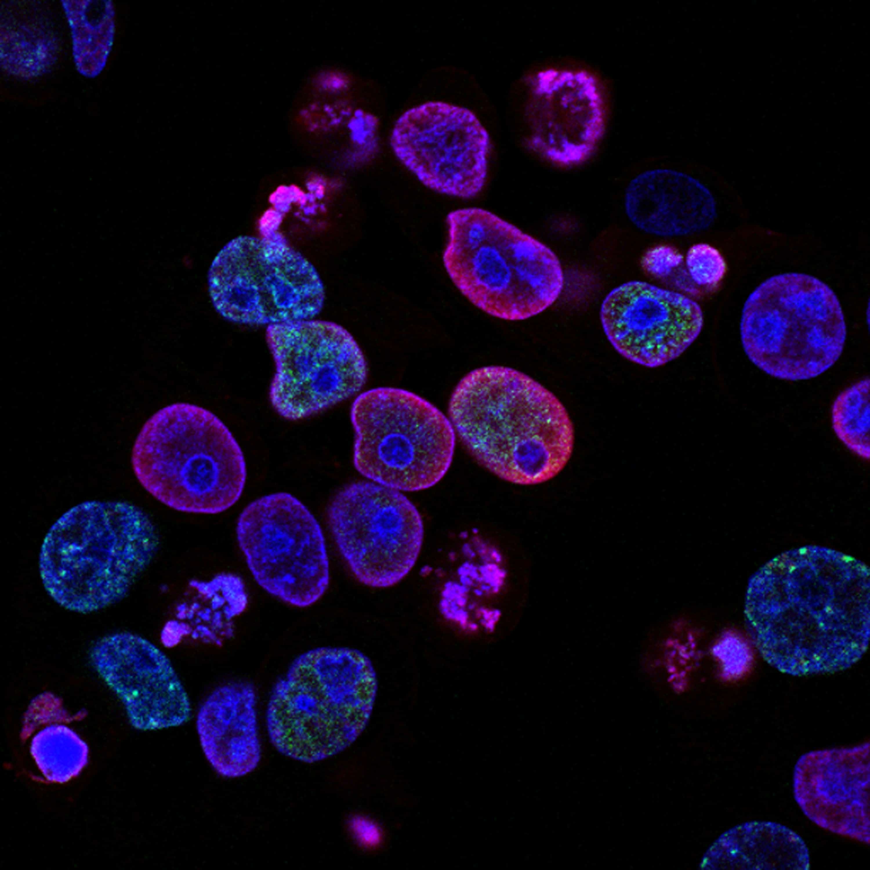
The PRODI team has been developing a new digital imaging method over the last years. The so-called label-free infrared (IR) imaging measures the genomic and proteomic composition of the examined tissue, i.e. provides molecular information based on the infrared spectra. This information is decoded with the help of artificial intelligence and displayed as false-colour images. To do this, the researchers use image analysis methods from the field of deep learning.
In cooperation with clinical partners, the PRODI team was able to show that the use of deep neural networks makes it possible to reliably determine the so-called microsatellite status, a prognostically and therapeutically relevant parameter, in colon cancer. In this process, the tissue sample goes through a standardised, user-independent, automated process and enables a spatially resolved differential classification of the tumour within one hour.

Effectiveness of therapies
In classical diagnostics, microsatellite status (the amount of abnormalities) is determined either by complex immunostaining of various proteins or by DNA analysis. “15 to 20 per cent of colon cancer patients show microsatellite instability in the tumour tissue,” says Professor Andrea Tannapfel, head of the Institute of Pathology at Ruhr University. “This instability is a positive biomarker indicating that immunotherapy will be effective.”
With the ever-improving therapy options, the fast and uncomplicated determination of such biomarkers is also becoming more and more important. Based on IR microscopic data, neuronal networks were modified, optimised, and trained at PRODI to establish label-free diagnostics. Unlike immunostaining, this approach does not require dyes and is significantly faster than DNA analysis. “We were able to show that the accuracy of IR imaging for determining microsatellite status comes close to the most common method used in the clinic, immunostaining,” says PhD student Stephanie Schörner. “Through constant further development and optimisation of the method, we expect a further increase in accuracy,” adds Dr. Frederik Großerüschkamp.
Selected for you!
Innovation Origins is the European platform for innovation news. In addition to the many reports from our own editors in 15 European countries, we select the most important press releases from reliable sources. This way you can stay up to date on what is happening in the world of innovation. Are you or do you know an organization that should not be missing from our list of selected sources? Then report to our editorial team.
Related Posts


