
If a suspicious lump is discovered in a patient’s breast, a biopsy is usually called for and a doctor will take small portions of tissue from the lump and have them examined in the laboratory. It is vital to find out whether the suspicious lump is a harmless change in the tissue or whether it is a sign of disease, i.e., a tumor. Currently, patients have to wait several days for the results. What’s more, a biopsy is expensive, painful and – because it is an invasive method – brings with it certain risks, such as the wound becoming infected or neighboring tissue becoming damaged, writes the Fraunhofer Institute in a press release.
Quick and gentle: screening biomarkers in bodily fluids
Scientists at the Fraunhofer Center for Applied Nanotechnology CAN, a research division of Fraunhofer IAP in Hamburg, have set themselves the goal of making the conventional biopsy used to diagnose breast cancer redundant – along with all the disadvantages it entails. “In the LIBIMEDOTS project, we are working on detecting breast cancer via circulating tumor cells – together with Spain’s Universitat Rovira i Virgili, Universität Hamburg and the Medical Center Hamburg-Eppendorf,” explains Dr. Neus Feliu Torres, who has been leading the “Nanocellular Interactions” working group at Fraunhofer IAP via an Attract program since July 2020.
If there is a tumor in the body, for example in the breast, a number of tumor cells may enter the bloodstream and other bodily fluids. If these cells can be detected there instead of by taking a biopsy of the tumor, there are numerous advantages. Firstly, the method is gentle — the doctor simply has to take a little blood from the patient. Secondly, the results are available within just a few hours. By pursuing this approach, the team of researchers hopes that, as well as discovering early-stage breast tumors faster and in a gentler manner, it will also be possible to assess whether a treatment is successful.
Enriching and detecting tumor cells
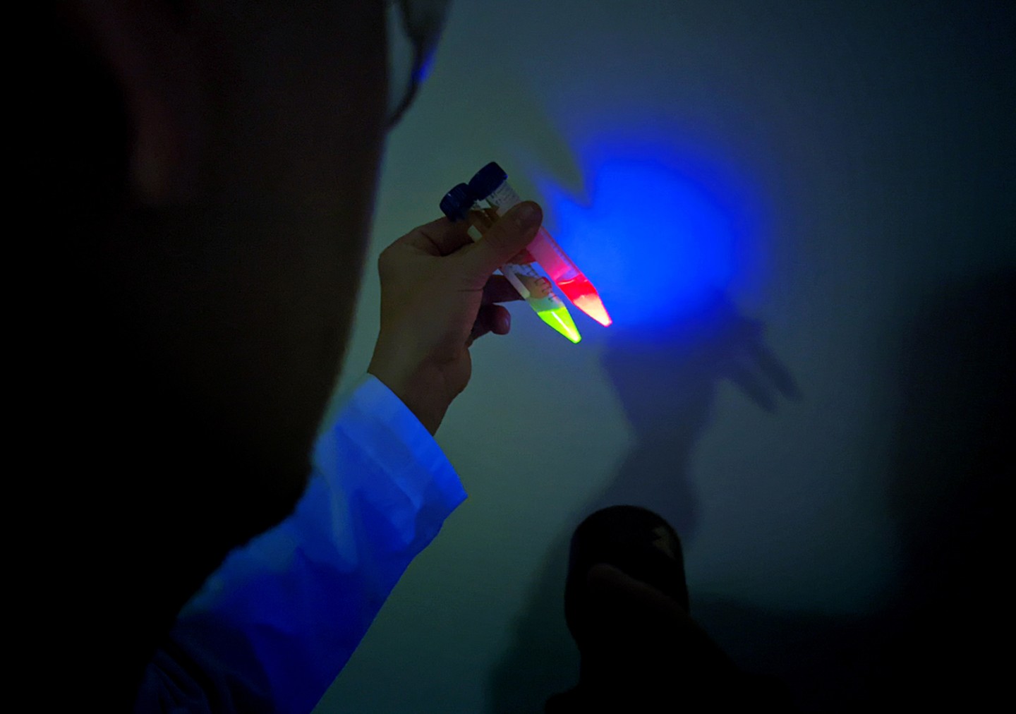
The examination principle is based on two pillars. The first involves enriching the tumor cells in the blood to enable them to be detected. “Compared to other somatic cells, tumor cells in the blood are only present at a ratio of one to one million. They are therefore extremely difficult to find and detect,” says Feliu Torres. To enrich the tumor cells, the Fraunhofer team is developing magnetic nanoparticles. This cargo enables the tumor cells to be collected and concentrated by a magnetic field. The second pillar is the specific binding of fluorescent particles to the surface of the enriched tumor cells in order to detect them. “This allows us to use the surface of the tumor cells for the specific binding of fluorescent particles. These then light up so brightly that just a few tumor cells need to be found to provide evidence,” explains the chemist and medical scientist.
As the circulating tumor cells vary from patient to patient, it is not enough to use just one type of fluorescent particle. The team is therefore developing a range of different particles with specific binding properties to uncover the tumor cells. This will make the detection process more sensitive, more specific and quicker, as well as more cost-effective.
Selected for you!
Innovation Origins is the European platform for innovation news. In addition to the many reports from our own editors in 15 European countries, we select the most important press releases from reliable sources. This way you can stay up to date on what is happening in the world of innovation. Are you or do you know an organization that should not be missing from our list of selected sources? Then report to our editorial team.