

Around three million animals, from mice to birds to fish to monkeys, cows or dogs, are used for animal experiments in science in Germany every year. Of that total, about 47 percent were used in basic research and about 13 percent in research on human and animal diseases in 2019, according to figures from the German Federal Ministry of Food and Agriculture. About 22 percent were needed in the production or quality control of medical products or for toxicological safety testing, and about 18 percent were used for other purposes such as education, training or for breeding genetically modified animals.
On February 8, 2021, the state of Baden-Württemberg established a statewide university network to improve animal welfare in research and teaching, funding it with €3.8 million. Future animal experiments are to be “avoided, reduced and improved.” One of the projects being financed is the 3R-US network of the University of Stuttgart and the Robert Bosch Hospital in Stuttgart. The two partners want to develop an ex-vivo tumor tissue platform to replace animal testing. The focus is on molecular diagnostics, biomaterials and simulation.
Step-by-step replacement of animal testing
Ultimately, a platform for cancer research should provide technologies and analytical methods “based on primary tumor tissue from humans to gradually replace animal testing.” A group led by Prof. Monilola Olayioye of the Institute of Cell Biology and Immunology (IZI) at the University of Stuttgart is investigating how signaling networks of oncogenes that promote tumor growth interact with tumor suppressors that inhibit it. Such mechanisms are responsible for the uncontrolled proliferation and spread of cancer cells. To stop this process and destroy the tumor, suitable sites of attack must be found where new active substances can dock that override these mechanisms.
“If we want to test such substances with as few test animals as possible, or eventually with no test animals at all, we have to reproduce the complex organism of a tumor as realistically as possible,” says Olayioye, describing the challenge. So far, he says, this has only been possible to a very limited extent. In tests with isolated cancer cells in a Petri dish in a nutrient solution, the potential active ingredient is fed directly to the cells. But this bears little resemblance to the normal distribution processes in the human body. And test systems in which human tumor cells or tissue are implanted in mice where they continue to grow are not the ultimate wisdom either, since they do not replicate the communication between cancer and immune cells in humans.

Ex vivo, de novo and in silico
Researchers are therefore looking for systems in which tumor cells can grow three-dimensionally and interact with other cell types. 3R-US builds on three approaches: ex vivo, de novo and in silico. In ex vivo experiments, drugs are tested outside the organism not on single cells but on tissue samples. However, since this tissue can only be used once even if continuously supplied with nutrients, the 3R-US team relies on so-called de novo technology. This involves using 3D printing techniques to create tissue-like structures from biomaterials and cells which are then cultured in tiny chambers (a microfluidic system) that allow for the controlled delivery of active ingredients.
“With the help of such microsystem technologies, we can assemble various biological structures like Lego bricks to replicate the corresponding tumor,” explains Michael Heymann, assistant professor at the Institute of Biomaterials and Biomolecular Systems at the University of Stuttgart. The data obtained in ex vivo and de novo tests will then be used to build and validate in silico models. This means that modeling and simulations, together with simulation experts, will enable more realistic predictions of drug distribution.
Cross-institutional 3R-US network
“If we are already able to filter out the best drug candidates in ex vivo culture systems, we can significantly reduce the number of experimental animals for the final preclinical tests,” says Prof. Roland Kontermann at IZI. “To replace animal testing altogether, de novo and in silico models will offer great potential in the future.”
Over the next five years, knowledge from the university and clinic will be combined in a cross-institutional 3R-US network. The goal is to create synergies between biotechnological and medical technology expertise and provide a service “that can also be used to sensitize students to the topic at an early stage.”
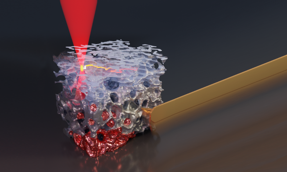
Cover photo: High-precision 3D printing can be used to create complex carrier structures (gray) and populate them with cells (red). The microstructure shown here is about as wide as a human hair. This technique will be used to replicate tumor tissues in great detail. © University of Stuttgart/IBBS
More articles on animal testing can be found here.
Related Posts


