
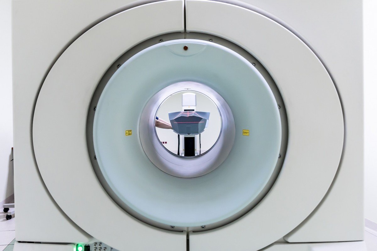
A constant buzz fills the space and in the middle of the room a large round tube with a hole in it. An MRI scanner. Today we’re taking a look behind the scenes at Scannexus in Maastricht. In this research centre, researchers, clinicians and entrepreneurs can find an answer to a question, from Alzheimer’s to knee examination. It’s all possible.
Scannexus assists with setting up an examination, processes data or brings in experts of other areas. Researchers work together with various research groups from Maastricht University or other European research centres. Job van den Hurk is a neuroscientist and data scientist at the research centre. He proudly discusses the MRI scanners in the building. Three in total, a 3 Tesla, 7 Tesla and 9.4 Tesla. (Tesla is a unit that indicates how strong a magnetic field is. For comparison: an ordinary refrigerator magnet is 0.005 tesla.) “Oh, and a dummy scanner. We use it to let children get used to lying still”, says van den Hurk. “Not very exciting otherwise”. He closes the door behind him and walks through the corridor towards his pride and joy. A 9.4 Tesla MRI scanner. “There are only about ten of these in the world. How spectacular is it that one of them is here?”
[learn_more caption=”How does MRI work?”] MRI stands for Magnetic Resonance Imaging, a magnetic field is generated according to the physical principle of induction where current is passed through wires in a coil. “These wires are connected to each other, so the current flows around. But this gives you resistance and a lot of heat. That would use so much energy that the whole South Limburg wouldn’t have electricity anymore, which is why we do it differently”, laughs Job van den Hurk. “When the coil is brought under a certain temperature, superconductivity is created, which means that the resistor completely disappears and the electrical voltage continues to flow around without the need for extra current. This generates the magnetic field. The coils are cooled with liquid helium – to -273 degrees Celsius – to uphold the magnetic field.
In the scanner, particles within the centres of hydrogen atoms (that are located everywhere in the body) react to the magnetic field of the MRI scanner. Just like the needle of a compass, the particles in the cores are focussing on the magnetic field. As we mostly consist out of water, this happens throughout the body. These magnetic vibrations are captured by another coil in the device. Then a computer makes a 3D recording of these vibrations. [/learn_more]
In the bicycle shed
It all started about six years ago in the bicycle shed of the faculty of psychology. “At first mainly for our own research, but more and more other requests came in. It has gradually developed into Scannexus. The old scanner is no longer there, it has been exchanged for a new version of 3 Tesla”, says van den Hurk. Then laughing: “Tesla has nothing to do with the car, but with the strength of the magnet. In hospitals, you will find scanners between 1.5T and 3T. The 1.5T is mainly used to diagnose damage to ligaments and tendons of joints. Brain scans in the hospital are usually done in a 3T scanner, on which you can see the difference between grey and white brain matter, with which you can diagnose diseases like MS.
Deeper into the brain
But if you want to dive deeper into the brain, you need 7T for this. “This is where it gets really interesting”, van den Hurk crawls behind a screen and opens a brain scan. “Look, with these kinds of images you can see all six outer layers of the brain. These images are far too complex for diagnosis and take up too much data. But that will change, as in Europe and America the scanners are now officially allowed to be used for diagnoses. Computers’ computing power is also increasing, which makes the operation and reading of images increasingly less complex. In a few years’ time, this will be the standard. Also, because MRI – unlike X-rays – have no harmful consequences for patients.
Van den Hurk opens the room that embraces the masterpiece of the research centre. A 50,0000-kilo magnetic tube fills the room. “The magnet is so strong that a cage with approximately 600 tons of steel had to be built around it. If the cage weren’t there, all the cars of the car park would come straight through the wall,” he says with a grin. This device makes images of the brain or spinal cord and has such a high resolution that researchers can see things on them that are normally only visible with a microscope. Van den Hurk: “The brain consists of layers and by combining high contrast and resolution, you can distinguish sub-layers from each other and look much deeper into the brain. It produces fascinating 3D images.”
The great advantage of such a strong scanner is that researchers no longer have to cut into tissue or examine a dead brain. “Cutting upon skulls to retrieve a microscope sample is outdated. Moreover, a living brain is much more interesting to look at as you can see structures that you normally don’t see. You can anticipate how groups of neurons communicate with each other and then get to compare them,” says van den Hurk.
Breaking walls
But it is not only the cool scanners’ that are important for the research centre. “The research questions that arise here are so complex that you need multiple disciplines to come up with a solution. What you used to see was that brain scientists had little knowledge of what for example biologists were doing. We try to break through these walls by working with several faculties or different researchers. This is how you learn from each other and how new ideas arise, to treat diseases for example.”
Currently, they are working on a study looking for an answer to the question why some people always have the feeling that they have to urinate. “How this exactly works in the brain is still unclear. We hope to find out by placing healthy people under the scanner and filling their bladder via a tube. They then press a button when they feel that their bladder is full, and researchers can see which areas in the brain then light up. The same process is then repeated with people who have bladder issues, to compare the results. By linking the data from this research to the expertise of urologists, we think we can find a solution faster,” says van den Hurk.
Related Posts


