

Three-dimensional imaging techniques are nothing new in medicine. They are familiar from conventional clinically-used computed tomography (CT). However, these images are still quite far from “normal” high-resolution images. Researchers at the Hannover Medical School (MHH), Mainz University Medical Center and the HELIOS University Hospital Wuppertal at the University of Witten/Herdecke, all in Germany, have now found a way to improve the results many times over.
They lead an international, multidisciplinary consortium that enables high-resolution, three-dimensional X-ray images of the human body. With a maximum resolution of up to 300 nanometers, they correspond to one-tenth the diameter of a human hair.
“The networking between radiology, pathology and molecular diagnostic approaches will assume enormous clinical importance in the future,” says pathologist Prof. Danny Jonigk, MHH Institute of Pathology. “With high-resolution three-dimensional imaging, we can better understand tissue damage to the lung in COVID-19,” adds his colleague Dr. Maximilian Ackermann from the Institute of Pathology at Helios University Hospital Wuppertal, University of Witten/Herdecke, and the Institute of Functional and Clinical Anatomy at the University Medical Center Mainz.
A hundred times more accurate than a conventional CT
High-resolution microcomputed tomography using synchrotron radiation is based on high-energy X-rays generated using synchrotron particle accelerators. The result is much higher resolution images. The scientists of the “Human Organ Project” from Germany, the European Synchrotron Facility ESRF in Grenoble, France, and bioinformatics and biomechanics experts from University College London (UCL), UK, have access to a recently reconstructed radiation source of the ESRF in Grenoble.
According to the researchers, this “Extremely Brilliant Source” (EBS) is the “world’s first fourth-generation high-energy synchrotron source and currently the brightest X-ray source in the world.” The project promises images of the entire human body with a resolution of two micrometers, more than a hundred times better than a CT scanner, they said. “We were very pleased that the Chan Zuckerberg Initiative is funding our translational approach,” explains Dr. Ackermann.
Focusing on tissue damage from COVID-19
This novel, high-resolution imaging technique will certainly provide fascinating new insights into other diseases, such as cancer or Alzheimer’s dementia, the researchers believe. In view of this, they are currently concentrating, together with colleagues in England and France, on spatially characterizing and understanding the tissue damage caused by COVID-19 pneumonia using this innovative method.
The project is funded by a $1 million grant from the Chan Zuckerberg Initiative (CZI) of Facebook founder Mark Zuckerberg and his wife, pediatrician Dr. Priscilla Chan. The initiative was launched in December 2015 in East Palo Alto by Mr. Zuckerberg and Dr. Chan to promote engineering, basic biomedical research and artificial intelligence.
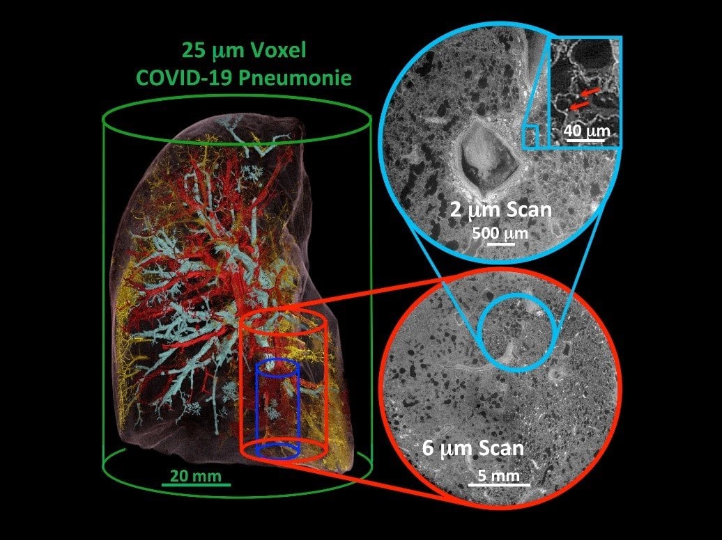
Cover photo: Three-dimensional segmentation and reconstruction of a lung with COVID-19 pneumonia. Individual thrombi (yellow), vascular changes (red) and airway changes (turquoise) can be visualized via the high-resolution method of Hierarchical Phase-Contrast Tomography (HiP-CT). Copyright: Institute of Pathology/MHH
Related Posts


