

Endoscopies, as in a gastroscopy or colonoscopy, produce images from the inside of the body. However, the instruments that make such images possible are usually at least as thick as a fingertip. This means that it is impossible to look inside blood vessels. That’s a real shame. Because endoscopies in veins could help prevent strokes or even heart attacks if deposits in the blood vessels are detected early on.
This possibility is now within arm’s reach thanks to fiber optic technology. These fibers – as thin as a hair – are only 125 micrometers thick. Nevertheless, it is still not that easy to develop optics that bend a laser beam to the side, scan the vascular wall, and direct the reflected light back to the fiberglass.
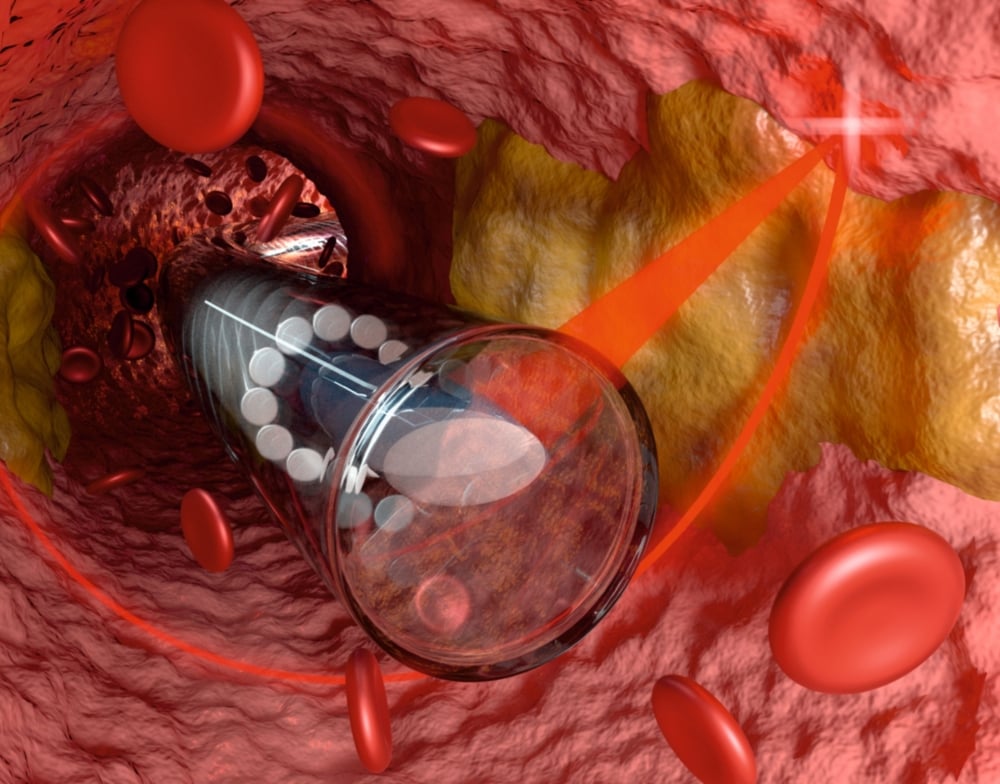
High-precision images in 3D
Researchers from the universities of Stuttgart (Germany) and Adelaide (Australia) and several other research institutes have now developed an optic instrument with a diameter of only 125 µm. This can be printed directly onto the fiberglass using an ultra-precise 3D printer. According to the researchers, these micro-optics are capable of diverting the laser light to the side, focusing it at a point “and at the same time correcting the distortion of the laser beam when it passes through a capillary-like plastic sheath with a diameter of less than 0.5 mm that has been fitted to protect the endoscope.” Here, the physician uses the laser beam to spirally scan the inside wall of a blood vessel. This allows the physician to obtain very precise 3-dimensional images – directly from inside the vein.

This smallest endoscopic optic system in the world has a diameter of less than half a millimeter, including the sheath. It was tested by researchers in Australia. They combined it with their OCT imaging equipment and then placed it in a human carotid artery and in the arteries of mice. The scientists discovered that “by rotating the optic system in that flexible mantle, they were able to take 3-dimensional images of the inside of the blood vessels in extremely high resolutions. During further examination of the blood vessels, they also discovered that they could detect both early-onset plaque deposits and cholesterol crystals (the main causes of vascular disease) in the contactless laser OCT endoscopy images.
Endoscope inside veins with an inner diameter of just 0.5 mm
Dr. Simon Thiele, who is responsible for the design of these miniature optic systems, is hoping that it will be possible to detect plaque deposits in time in the future. “And perhaps one day it will be possible to dissolve these thrombi in time by using a suitable laser beam,” the Stuttgart scientist said.
The goal now is to commercialize the 3D printed mini-optic systems as part of a spin-off. The company Nanoscribe GmbH from Karlsruhe (which was itself a spin-off company from the German Karlsruhe Institute of Technology – KIT -11 years ago) built the ultra-precise 3D printer. Carl Zeiss AG, based in Oberkochen, the world market leader in medical technology, already has a stake in Nanoscribe. The researchers were backed by the German Federal Ministry of Education and Research, the Baden-Württemberg Foundation and the German Academic Exchange Service (DAAD).
Main photo: Miniature optics in a vein. ©Florian Sterl, Sterltech Optics
Related Posts


