
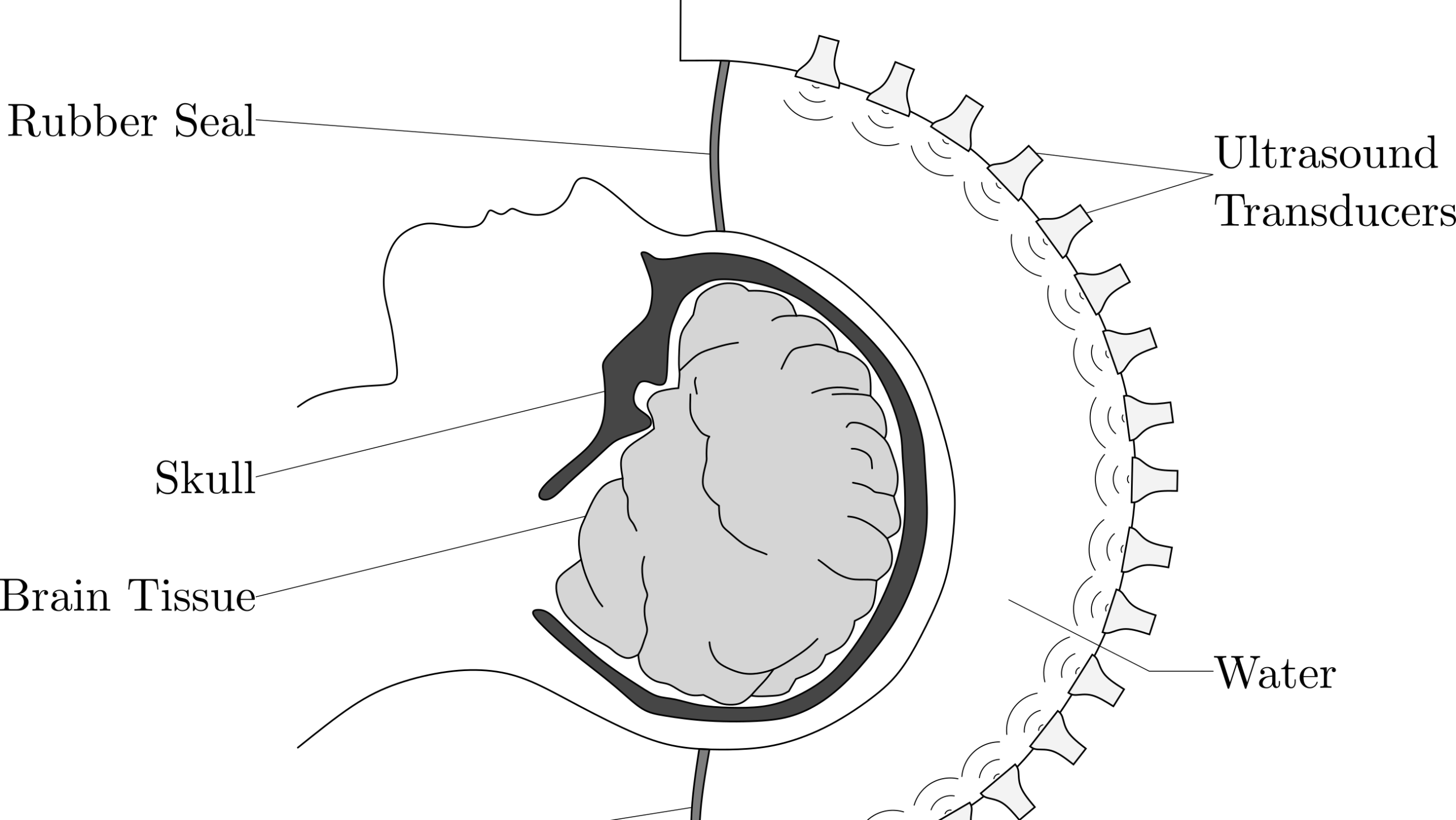
Researchers from ETH Zürich have developed a technology based on ultrasound to examine the human body non-invasively and cost-effectively. This method could provide the basis for imaging the brain with ultrasound in high resolution. Compared to computed tomography (CT) or X-rays, ultrasound has a decisive advantage, it is harmless to the body. This makes it possible to determine the tissue type and distinguish whether it is a brain mass or tumor tissue, for example. Moreover, it is much more cost-efficient than magnetic resonance imaging (MRI), writes ETH Zürich in a press release.
Waves
The technology is developed on the basis of ultrasound for medical imaging and seismology for imaging the Earth’s interior to measure the propagation of waves through matter. For instance, when seismic waves encounter material differences in the Earth’s interior, they are reflected and refracted at their interfaces. As a result, the speed of the waves changes. If researchers measure these waves at the surface, they can draw conclusions about the structure of the Earth’s interior.
With the help of sophisticated algorithms and high-performance computers, professors can use this wave data to characterize the three-dimensional structure of the Earth. The parallels to propagation between ultrasound and earthquake waves, as well as the team’s know-how in the field of wave physics, led the ETH scientists to also study wave propagation for medical ultrasound.

Learning on a magnetic resonance imaging scanner
For their model, the researchers first use an MRI of the brain as a reference. Then they perform calculations with different parameters until the simulated image matches the MRI’s.
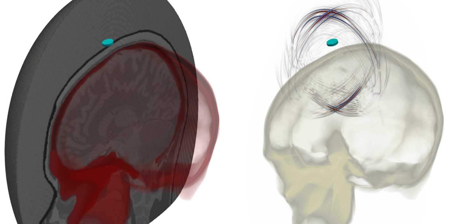
This method obtains a quantitative image instead of the less informative greyscale image common to conventional ultrasound. By using all the information from the complete wave field, the researchers can correctly map the physical properties of the medium at every point in the brain.
A particular remaining challenge is the complex geometry of the skull, due to eye, nose, and jaw cavities, which must be modeled precisely in the simulation without dramatically increasing the computation time. To solve this problem, Marty is developing methods to create individual numerical meshes for arbitrary skull shapes out of hexahedra (small elements with six faces).
Selected for you!
Innovation Origins is the European platform for innovation news. In addition to the many reports from our own editors in 15 European countries, we select the most important press releases from reliable sources. This way you can stay up to date on what is happening in the world of innovation. Are you or do you know an organization that should not be missing from our list of selected sources? Then report to our editorial team.


