
Scientists have introduced a groundbreaking technique called Embedded Extrusion-Volumetric Printing (EmVP). This technique overcomes challenges in bioprinting and create physiologically-relevant models. As a proof-of-concept, EmVP has been applied to create synthetic biology-inspired intercellular communication models. The research team published their findings in Advanced Materials.
EmVP: A novel technique for bioprinting complex structures
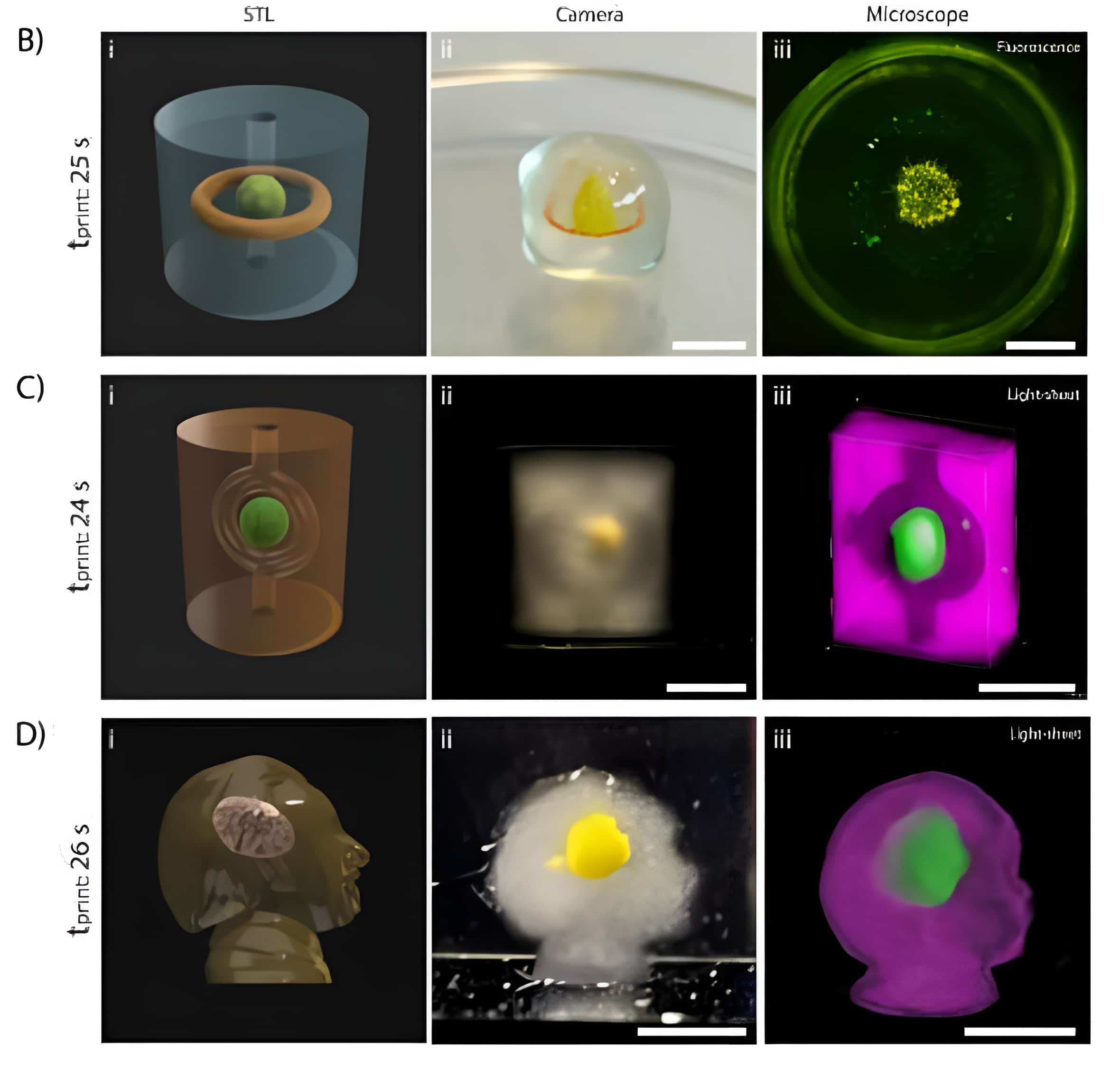
The EmVP technique combines extrusion-bioprinting and layer-less, ultra-fast volumetric bioprinting, allowing the spatial arrangement of multiple inks or cell types. This innovative method addresses the limitation of accurately replicating the intricate structures and patterns of cells at both the micro- and macroscale, which has hindered the creation of models that closely resemble physiological conditions.
Developed by an international team of researchers mainly from Utrecht University, EmVP has the potential to revolutionise bioprinting and its applications in medical research and other industries. The technique has been successfully applied to create complex synthetic biology-inspired intercellular communication models, providing new avenues for producing regenerative grafts, developing engineered living systems, and advancing (metabolic) disease models.
EmVP’s potential in medical research: Parkinson’s disease
One specific application of the EmVP technique is the development of an in vitro model of a neural network, as demonstrated in the thesis of Dieuwke De Vos, one of the researchers involved in EmVP. De Vos and her team used suspended extrusion-based bioprinting to establish a 3D in vitro model of a neural network for studying Parkinson’s disease, a condition that affects an increasing number of people worldwide and causes central nervous system cells to malfunction, resulting in tremors, stiffness, balancing issues, and rigidity.
Traditional models for studying Parkinson’s disease include simplified 2D models and animal models, both of which have limitations in replicating human disease. The EmVP technique allows for the printing of 3D human brain tissue for studying Parkinson’s disease, providing a more accurate representation of the affected tissue and offering new opportunities for drug testing and personalised medicine applications.
How EmVP works: Bioresins, microgels, and support baths
To develop the EmVP technique, the research team created light-responsive microgels to be used as bioresins (µResins) for light-based volumetric bioprinting. These µResins provide a microporous environment for cell homing and self-organisation, enhancing the differentiation of multiple stem/progenitor cells, including vascular, mesenchymal, and neural cells.
By tuning the mechanical and optical properties of gelatin-based microparticles, the researchers were able to use them as a support bath for suspended extrusion printing. This process involves printing bioinks containing living cells into a support bath, which prevents the collapse of the printed structure and maintains cell positions. The EmVP technique allows for the rapid creation of high-resolution features containing multiple inks and cell types, offering advantages over conventional bioprinting methods.
Future prospects for EmVP in bioprinting and beyond
The introduction of the Embedded Extrusion-Volumetric Printing (EmVP) technique marks a significant step forward in bioprinting technology, enabling the creation of complex 3D structures with multiple inks and cell types. Its potential applications in producing regenerative grafts with biological functionality and developing engineered living systems and (metabolic) disease models open up new possibilities for medical research and the bioprinting industry as a whole.
As EmVP continues to be refined and applied to various research areas, its impact on the study of diseases like Parkinson’s and the development of personalised medicine is expected to grow. The technique’s ability to create physiologically-relevant models may lead to significant breakthroughs in understanding and treating various medical conditions, ultimately improving patient outcomes and quality of life.
Related Posts


