

For a long time, microglial cells were thought to be immune cells in the brain that were mediocre performers. It wasn’t until the early 1990s that it was discovered that they actively shape our nervous system. In the first years of life, they sever weak connections to fine-tune the network of nerve cells. This is how perceptions become permanently anchored in the brain.
The network that connects nerve cells and stores memories is called the perineuronal network. It is especially adaptable at this early stage of development and considerably freer than later on in adulthood..
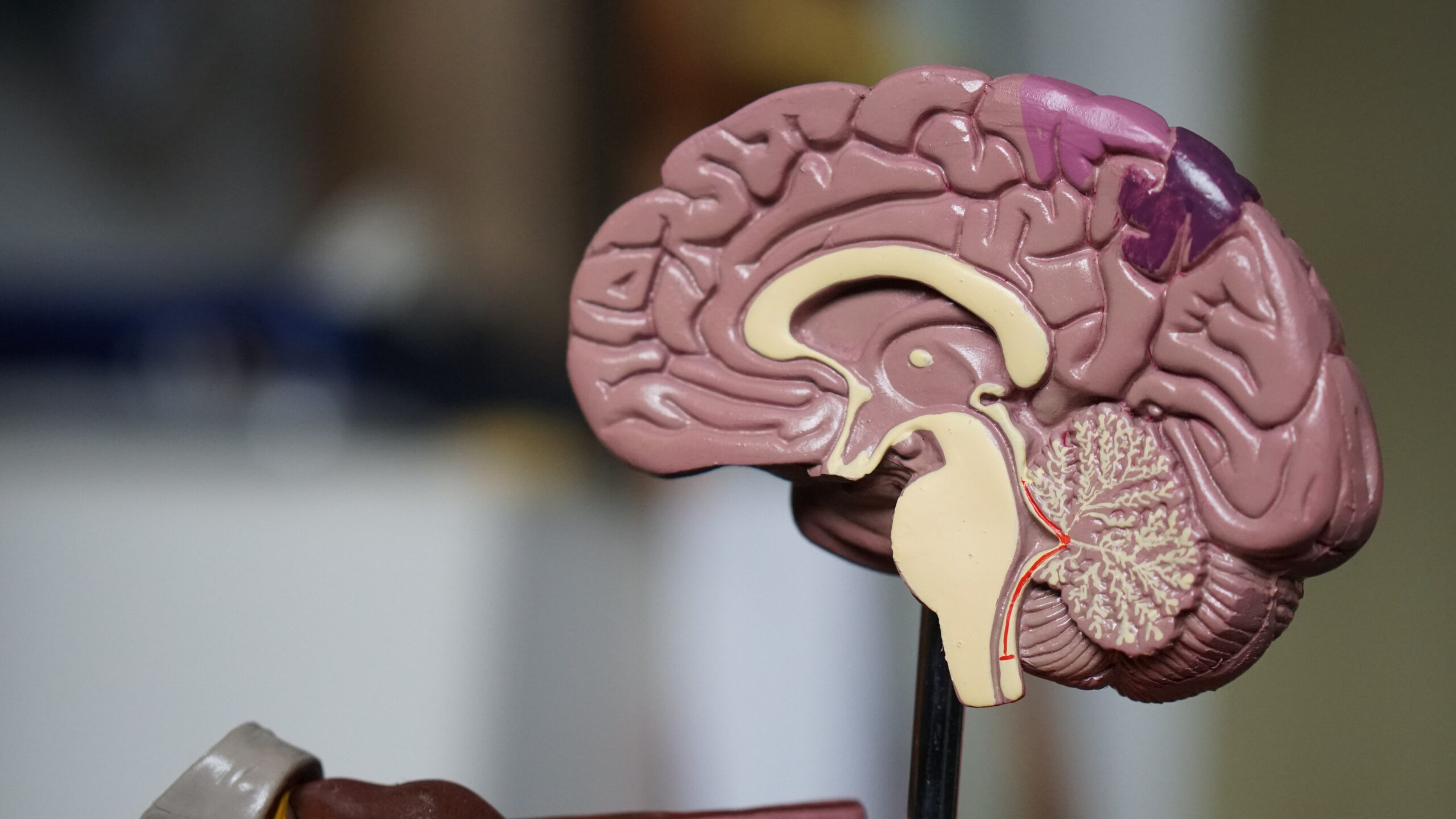
Microglia cells
During the early childhood phase, microglial cells are in a reactive state. Later, they take on an observer role. The transition is fluid, but is also evident in the shape of the cells. In the observer mode, microglia run like roots through the healthy brain and control its functions. Reactive microglia take on a spherical shape and surround the tissue that they are breaking down.
Ketamin
Neuroscientist Professor Sandra Siegert, who teaches and conducts research at the Institute of Science and Technology (IST), Austria, wants to understand the role of microglia in the healthy and diseased brain in order to come up with new therapeutic treatments. As early as four years ago, an experiment by her team led to a sensational result: The researchers anesthetized mice with ketamine, a key drug in surgery that has also recently been approved for use in psychiatry. In the process, they discovered that microglial cells in the mice’s brains became highly reactive.
The researchers knew that microglial cells can also eat synapses and whole neurons. This is a phenomenon that often occurs in the late stages of Alzheimer’s disease. So, at first, only the strength of the reaction surprised them. But when they saw that the microglial cells were eating the perineuronal network, and not the neurons and synapses as had been expected, their enthusiasm erupted. A significant loss of the perineuronal network was observed after only three treatments. As a result, the blockages dissipated and it network is again receptive to new inputs and synapses.
Light therapy
The researchers recently applied another procedure that generated a lot of excitement once again: they treated the mice with light that flickered 60 times per second and managed to unblock the perineuronal network this way as well. An effect that can be explained by now neurons communicate, which send electrical impulses to each other. These impulses become coordinated in such a way that waves of signals are created – so-called brain waves.
These waves can be affected by external sensory impressions, for instance by shining light into the eyes. Consequently, the researchers discovered that a precise coordination exists between different brain waves and the activity of microglia. These results could form the basis for a therapeutic treatment that could be applied to humans. By restoring the brain’s plasticity, they could potentially override traumatic experiences and treat post-traumatic stress disorder. “But we are very cautious, because something traumatic could also happen during this formation window,” the neuroscientist cautions.
Overwriting traumas
The research is still in its infancy. At the moment, researchers are conducting behavioral studies on mice to see if light therapy is linked to negative side-effects. Siegert: “So far, the opposite seems to be the case. The mice seem to perform better. Therefore, we are researching whether a certain duration of stimulation and a certain number of repetitions determine beneficial, as well as any potential detrimental effects.” Then there is the matter of finding out where certain memories are stored. Often though, a complex network of neural circuits sprawls across many different brain regions. By tuning into a specific frequency, it might become possible to modulate the entire network of interconnected neurons.
The researchers are also still speculating about the types of potential therapeutic treatments. “There might be several ways to combine light therapies with behavioral therapy. We think that we will see only beneficial effects with this combination.” The tool could work like a daylight lamp or light therapy glasses. But there are other potential uses for these treatments. One is amblyopia, a visual disorder caused by an imbalance of visual input during childhood development. If left untreated, it leads to permanent vision loss.
Another area that the researchers plan to explore are the molecular mechanisms behind their discovery, which are not yet fully understood. What is known for certain is that the methods have an advantage over existing approaches aimed at removing the perineuronal network. Both 60-hertz light flickering and ketamine treatment are simple and minimally invasive. In contrast, enzymatic removal with the chondroitinase ABC enzyme is extremely invasive and time consuming. It is a strategy with some limitations that stand in the way of therapeutic application, Siegert explains. To begin with, the enzyme must be injected directly into the brain, because it cannot cross the blood-brain barrier. There, it breaks down the entire extracellular matrix and not just specifically the perineuronal network. Moreover, the breakdown of the extracellular matrix takes several months, which could cause the brain to become hypersensitive.
Also interesting: Blood tests can detect Alzheimer’s disease at an early stage
Publication
The results of the study was published in the professional journal Cell Reports: Microglia enable mature perineuronal nets disassembly upon anesthetic ketamine exposure or 60-Hz light entrainment in the healthy brain.