

One challenge when it comes to optical imaging is the ability to get a good look at the inside human tissue. Up until now, imaging was usually couldn’t go further than a millimeter. Researchers from Delft University of Technology (The Netherlands) have been able to increase this to four times that depth when they study zebrafish. This breakthrough, which was ten years in the making, can visualize tumors more clearly.
“It’s basically about how deep you can look into tissue and being able to see its structure,” explains Jeroen Kalkman, speaking on behalf of the Delft research team. “The main technique we rely on is optical coherence tomography, OCT. This is already fairly old, it has been used since 1991 and is still successfully used in ophthalmology. OCT involves making a scan of the structures of the eye. We wanted to see how much further we could get. Whether we could get a deeper view, more in depth imaging.”
OCT is used by ophthalmologists to visualize the retina. Plenty of commercial parties already make OCT equipment. OCT is similar to acoustic ultrasound, but it uses light instead of sound waves to obtain a higher resolution.
Looking into the fog
Optical imaging, deep in the tissue, is limited due to light dispersion. You can compare it to looking into a fog, Kalkman explains. This dispersion reduces the depth of the image, the contrast, as well as the spatial resolution. Consequently, one way to be able to penetrate further optically is to remove that dispersion.
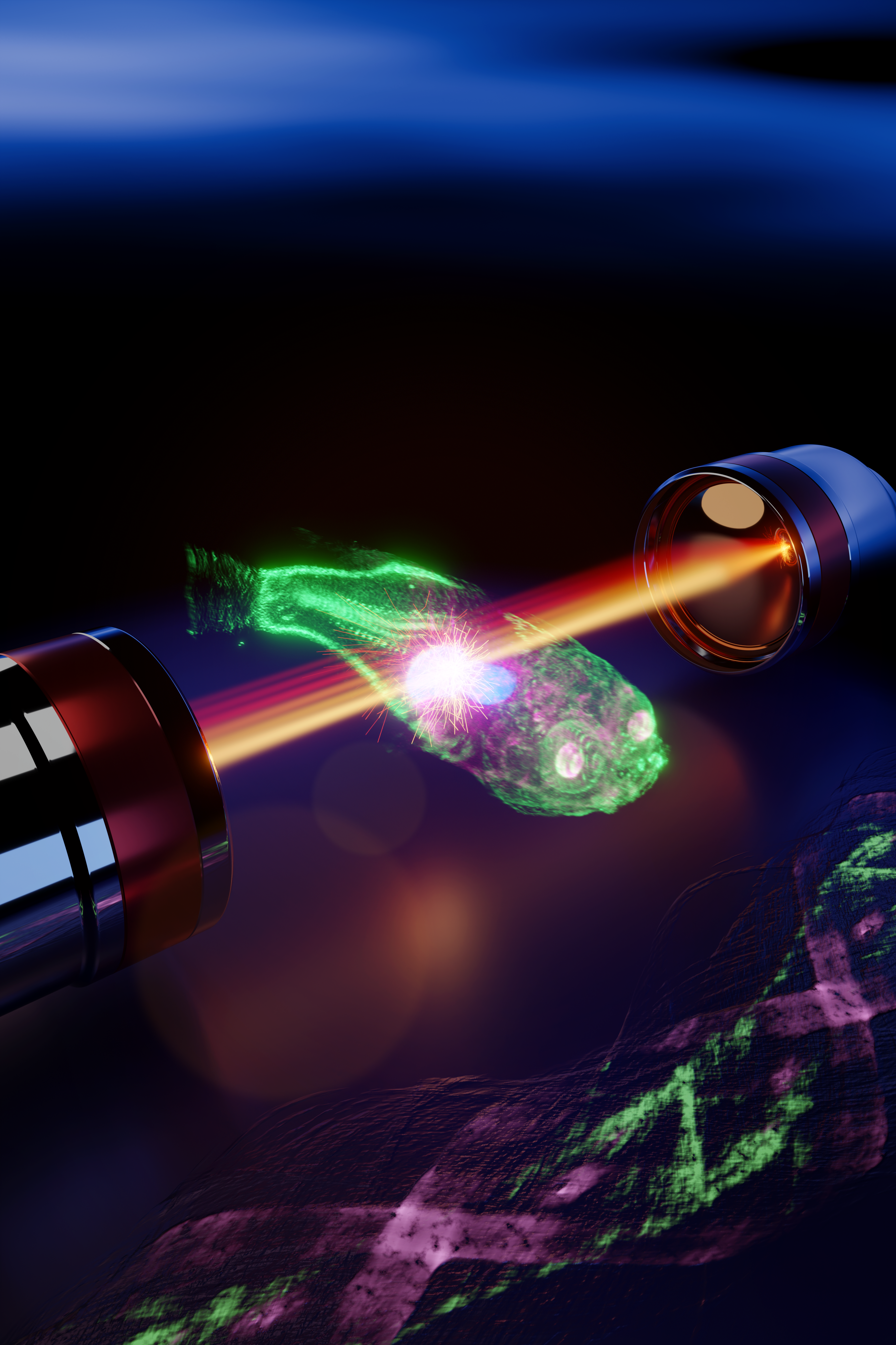
The method used to see more deeply into tissue involves a modification of the existing OCT technique. “Instead of reflecting light, as is customary, we send the light through the tissue. You capture the transmitted light. If you look at the light that is coming straight towards you, then you know where it has come from. But if the light has been scattered, as is the case when you look into a fog because of all the droplets, then you don’t know where it has been and you don’t get a sharp picture.”
Capturing this light is done using a sensor that’s placed on the other side of the tissue. It can be used to determine how fast and which light arrives. “The fog effect can be eliminated and sharp images can be obtained by separating the ‘fast’ light, the light that comes straight towards you, from the ‘slow’ light, which is slightly scattered.”

Just like an x-ray CT scan
One disadvantage of measuring with transmitted rather than reflected light is that no direct 3D spatial information is obtained. The Delft research group has found a solution to this issue with the use of computerised tomography.
“Computerized tomography is best known for its X-ray CT scan. But instead of X-rays, a 3D image is now created with a computer using visible light.”
“We use a similar algorithm and that creates a cross section of the object out of the information from the transmitted light waves. Unlike reflection, this algorithm now uses all images measured from all angles at the same time. Which makes imaging a lot more complicated.”
Testing it out on aquarium fish was a logical choice. “We work with zebrafish because they are popular among biologists as models to do experiments with. We managed to get aquarium animals through cooperation with the Erasmus MC university hospital, where they are used for ongoing research.”
Development of tumors and biopsies
For instance, zebrafish are widely used in disease research. “This technique allows you to better monitor the development of diseases. For one thing, you are able to monitor the effects of drugs on the development of tumors better thanks to the improvement in the visibility of tissue.”
“The other advancement is the usefulness of three-dimensional imaging in biopsies. Biopsies are small pieces of human tissue that medical practitioners use for diagnosis. Current practices can potentially become faster and more accurate with the Delft approach.”
It took ten years from the earliest days to the recent publication of the Delft method in Optica. Kalkman: “That’s the time span between the idea for improving the technique until now. It has taken quite a while for all kinds of reasons, like being able to secure funding and solve specific questions.”
The next step is to automate, speed up the method, apply it practically and manage to “sell” it. Kalkman explains what they mean by that. “It’s a fundamental innovation that at the moment still needs to be converted into a practical application. It’s not just about imaging techniques. Users such as pharmacists must also be willing to make the switch.”
Pharmacists have to want to make the switch
“Such as switching to using fish instead of mice. That step is difficult if they are used to mice. Getting them to accept that it is important for contemporary research. It’s often difficult when it comes to applications to try something fundamentally new. It’s a bit like the chicken and the egg dilemma, when they don’t know if it works. You really have to go public with it as a research group.”
The four-millimeter ‘deep’ imaging that was achieved was not an absolute must, but more of a motivation to see how far TU Delft’s research could go. “The motivation was really about pushing the limit, seeing what is possible. Compare it to top-level sports. We won’t be able to improve on that four millimeters all that much, I’m pretty convinced of that.”
Related Posts


