

Artificial intelligence is taking on more and more tasks in our modern world. For example, we use it every day when we use online search engines. Translation programs are unimaginable without AI, as are speech recognition, face recognition, computer games and, in the future, autonomous driving. In medicine, AI is also becoming more widespread and has already found its way into the operating theater. Just a few days ago, Innovation Origins wrote about operating with live 3D image navigation inside the body.
The Karlsruhe Institute of Technology (KIT) has now gone one step further and has even been awarded the NEO 2019 Innovation Prize (worth €20,000) by the Karlsruhe TechnologyRegion for their ‘HoloMed’ system. The new system assists surgeons in the operating room via Artificial Intelligence (AI) and Augmented Reality (AR). It does this by creating a model from computer tomographic images of the patient. These reveal the hidden structures deep inside the body.
GPS for the brain
HoloMed’s main focus is on cranial punctures. This is a procedure whereby accumulated fluid is removed from the brain in order to reduce pressure. Frequently used for e.g. brain hemorrhages, craniocerebral trauma and strokes. In order to determine the optimal point of insertion and alignment for the puncture, the surgeon must measure and glean data from “various anatomical landmarks” from computer tomography (CT) and/or magnetic resonance imaging (MRI) scans.
“The difficulty lies in the fact that determining the angle of insertion only allows for a very small margin of error and the doctor isn’t able to see the target straightaway,” notes Professor Björn Hein. He oversees the project together with Professor Franziska Mathis-Ullrich at KIT. Determining this exact point is complicated as these images are only two-dimensional and the human head is three-dimensional. That’s why only about 60 percent of all free-hand incisions are able to pinpoint the best position.
Surgeons use HoloMed augmented reality glasses to assist them in determining this optimal insertion point and angle for the puncture needle. An AI developed at the AI by science staff member Christian Kunz uses the data from the patient’s digital file and their latest CT and/or MRI scans for creating a model that accurately depicts the structures deep inside the body that cannot be seen externally. This information is superimposed onto the surgeon’s AR glasses and shows the surgeon precisely where and how to guide the needle, much like a navigation system.
Easy to use and cost-efficient
Professor Hein states that machine learning methods are used in the automated generation of this information. “First of all, a segmented 3D model of the head is generated, which is used to determine the target position. However, the doctor is always able to make their own adjustments if appropriate,” Hein adds. The aim of the system is to provide an “innovative, novel and cost-effective solution that has a direct influence on the quality of these procedures”.
After its puncture method is successfully rolled out, HoloMed will also be used for other operations in the future. Since the system is, firstly, easy to use, and secondly, cost-efficient, the inventors say it is ideal for lowering healthcare costs. Plus it would also benefit poorly financed hospitals in emerging countries.
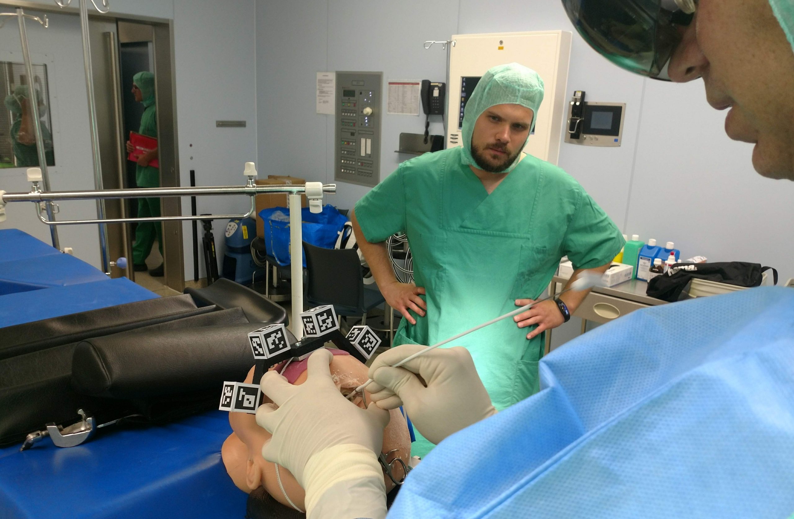
Cover photo: Dr. Michal Hlavac from the University Clinic for Neurosurgery Ulm and Christian Kunz from the “Health Robotics and Automation” (HERA) KIT team evaluating the HoloMed system during the initial surgery simulation with a dummy. (Photo: KIT-HERA).
Related Posts


